Blood Vessels Labeled Brain : Blood Vessels of the Brain Medical Illustration Medivisuals : Blood vessels are referred to collectively as the vascular system and, together with the heart, make up the circulatory system or cardiovascular system.
Blood Vessels Labeled Brain : Blood Vessels of the Brain Medical Illustration Medivisuals : Blood vessels are referred to collectively as the vascular system and, together with the heart, make up the circulatory system or cardiovascular system.. The blood vessels are the components of the circulatory system that transport blood throughout the human body. The brain and its surrounding blood vessels exist in a close relationship. The blood vessel wall is endowed with connective tissue, smooth muscle, and striated muscles. They bleed easily, which may cause epileptic attack, neurological problems and stroke. Blood vessels are intricate networks of hollow tubes that transport blood throughout the entire body so that it can deliver valuable nutrients to and remove waste from cells.
Blood vessels are referred to collectively as the vascular system and, together with the heart, make up the circulatory system or cardiovascular system. Cerebral arterial circle anterior communicating posterior cerebral a middle cerebral al reset zoom. Blood vessel endothelium is continuous with the inner tissue lining of organs such as the brain, lungs, skin, and heart. In the article on the ventricles within the cns, we will discuss their structure and. Only some of the vessels that exist in a real brain have been labeled.
MGH Martinos Center on in 2020 | Brain art, Anatomy art ... from i.pinimg.com Comes off the subclavian a., ascends although the internal carotid a. The capillaries also connect the branches of arteries and to. Capillaries surround body cells and tissues to deliver and absorb oxygen, nutrients, and other substances. Another whole article within the blood vessels and csf section is dedicated to the cavernous sinus. Supplies the anterior brain and the vertebral a. They bleed easily, which may cause epileptic attack, neurological problems and stroke. Brain vessel segmentation is a fundamental component of cerebral disease screening systems. Posterior communicating a internal carotid а.
Arteries transport blood away from the heart.
Blood vessels can be damaged by the effects of high blood glucose levels and this can in turn cause damage to organs, such as the heart and eyes, if significant blood vessel damage is sustained. The two cell types ensure the integrity of the neural vasculature by maintaining the blood. Blood vessels are vital for the body and play a key role in diabetes helping to transport glucose and insulin. The blood vessels (and nerves) enter the brain through holes in the skull called foramina. They bleed easily, which may cause epileptic attack, neurological problems and stroke. Arteries transport blood away from the heart. Learn vocabulary, terms and more with flashcards, games and other study tools. Blood vessels are referred to collectively as the vascular system and, together with the heart, make up the circulatory system or cardiovascular system. Only some of the vessels that exist in a real brain have been labeled. This is particularly important structure due to its clinical implications, which are discussed in more detail in the article. Microscopically, it is formed by the endothelium of the blood vessel. Blood is also supplied to the brain by the vertebral a. Blood travels from the heart in arteries, which branch into smaller and smaller vessels, eventually becoming arterioles.
Blood vessel endothelium is continuous with the inner tissue lining of organs such as the brain, lungs, skin, and heart. Cerebral arterial circle anterior communicating posterior cerebral a middle cerebral al reset zoom. The two cell types ensure the integrity of the neural vasculature by maintaining the blood. Blood vessels are intricate networks of hollow tubes that transport blood throughout the entire body so that it can deliver valuable nutrients to and remove waste from cells. Comes off the subclavian a., ascends although the internal carotid a.
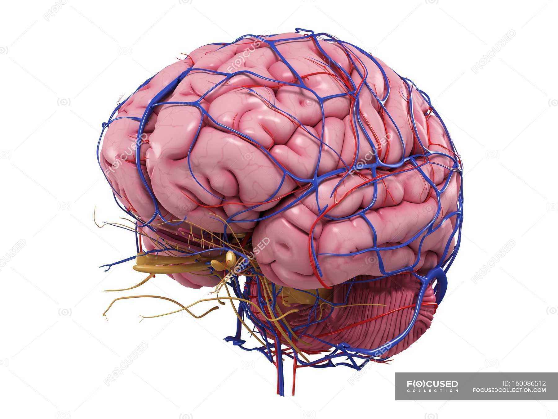
Human brain with network of blood vessels — circulatory ... from st.focusedcollection.com Cerebral arterial circle anterior communicating posterior cerebral a middle cerebral al reset zoom. The capillaries also connect the branches of arteries and to. Blood vessels in red in close communication with proliferating neuronal cells in the mouse cortex at embryonic day 10. • identification of blood vessels as arteries, capillaries or veins from the structure of their walls. Learn vocabulary, terms and more with flashcards, games and other study tools. The blood vessel wall is endowed with connective tissue, smooth muscle, and striated muscles. .blood vessels video, human body blood vessels, inner body, blood vessels labeled diagram, blood vessels labeling exercises, cat blood vessels labeled heart location, brain diagram labeled, digestive system diagram labeled, heart diagram labeled blood flow, heart diagram labeled detailed. Blood travels from the heart in arteries, which branch into smaller and smaller vessels, eventually becoming arterioles.
In the article on the ventricles within the cns, we will discuss their structure and.
Using medaka ( oryzias latipes ) as a model, the current protocol presents a quick and direct technique to label blood vessels in brain and pituitary by. The blood vessels are the components of the circulatory system that transport blood throughout the human body. There is a right sided aca and a left sided aca. Endothelial cells are labeled in red and pericytes in green. Fill in the blanks with the appropriate words to describe blood flow from the heart. Blood vessels in red in close communication with proliferating neuronal cells in the mouse cortex at embryonic day 10. The blood vessels (and nerves) enter the brain through holes in the skull called foramina. The difference in the structural characteristics of arteries, capillaries and veins is attributable to their respective functions. The blood vessel wall is endowed with connective tissue, smooth muscle, and striated muscles. The two cell types ensure the integrity of the neural vasculature by maintaining the blood. This is particularly important structure due to its clinical implications, which are discussed in more detail in the article. Capillaries surround body cells and tissues to deliver and absorb oxygen, nutrients, and other substances. Supplies the anterior brain and the vertebral a.
Fill in the blanks with the appropriate words to describe blood flow from the heart. Internal carotid artery (anterior circulation), vertebral artery (posterior circulation), and their hexagonal anastomotic network called blood brain barrier refers to the wall between the brain tissue and blood vessels. The capillaries also connect the branches of arteries and to. The structure, distribution and labeling of the whole brain vascular system of different arteries and veins in 3d. They also take waste and carbon dioxide away from the tissues.
Human Heart Labeled . Human Heart Labeled Heart Anatomy ... from i.pinimg.com Blood in the brain is supplied by two pairs of large blood vessels (arteries): The carotid arteries and the vertebral arteries anterior cerebral artery (aca): In the cerebral medulla, the arteries and veins of the right side of the body are controlled from the left side of the brain; Blood vessels flow blood throughout the body. Blood vessels can be damaged by the effects of high blood glucose levels and this can in turn cause damage to organs, such as the heart and eyes, if significant blood vessel damage is sustained. Cerebral arterial circle anterior communicating posterior cerebral a middle cerebral al reset zoom. The difference in the structural characteristics of arteries, capillaries and veins is attributable to their respective functions. The blood vessels are the components of the circulatory system that transport blood throughout the human body.
He says the restricted vessels prevent the blood from draining fast enough and injure the brain by causing a build up of iron which leads to ms.
Label the blood vessels of the male pelvis using the hints provided. Internal carotid artery (anterior circulation), vertebral artery (posterior circulation), and their hexagonal anastomotic network called blood brain barrier refers to the wall between the brain tissue and blood vessels. • identification of blood vessels as arteries, capillaries or veins from the structure of their walls. Cerebral arterial circle anterior communicating posterior cerebral a middle cerebral al reset zoom. Endothelial cells are labeled in red and pericytes in green. Supplies the anterior brain and the vertebral a. This vessel supplies blood to the front part of your brain, knows as your frontal lobe. Blood in the brain is supplied by two pairs of large blood vessels (arteries): The 500 ms patients, both adults and children, also underwent mri scans of the brain to measure iron deposits in surrounding areas of the brain. .blood vessels video, human body blood vessels, inner body, blood vessels labeled diagram, blood vessels labeling exercises, cat blood vessels labeled heart location, brain diagram labeled, digestive system diagram labeled, heart diagram labeled blood flow, heart diagram labeled detailed. Blood vessels are vital for the body and play a key role in diabetes helping to transport glucose and insulin. There is a right sided aca and a left sided aca. The structure, distribution and labeling of the whole brain vascular system of different arteries and veins in 3d.
The capillaries also connect the branches of arteries and to blood vessels labeled. Capillaries surround body cells and tissues to deliver and absorb oxygen, nutrients, and other substances.


0 Komentar